A 3D scanner that can help to diagnose heart disease or ischemia in under 2 minutes? It’s possible…
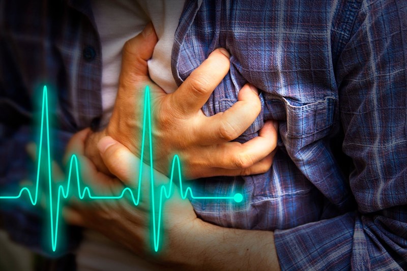
Chest pain… Cardiovascular disease… A leading cause of death around the world. According to the World Health Organization, more of the globe’s population die from cardiovascular causes than any other health condition. (1)
Classified as a group of disorders, cardiovascular diseases affect the heart and the body’s blood vessels (arteries, veins and capillaries) – the tubular structures which carry oxygenated blood to organs and tissues all around the body. A failure in this vitally important system can quickly become life-threatening. This group of diseases includes the likes of:
- Coronary heart disease (disease of the coronary arteries causing damage to the heart muscle)
- Cerebrovascular disease (an interruption in blood supply causing damage to the brain)
- Peripheral arterial disease (a circulatory problem involving the constriction / narrowing of blood vessels causing a reduction in blood flow to the limbs)
- Rheumatic heart disease (cardiac inflammation and scarring damage that occurs to one or more of the heart valves / heart muscle following a bout of rheumatic fever)
- Congenital heart disease (heart structure malformations which exist at birth)
- Deep vein thrombosis and pulmonary embolism (the formation of blood clots which can dislodge and travel through the circulatory system to the heart, lungs or even the brain)
Chest pain is taken seriously in any instance, mostly due to the possibility of a heart attack or stroke which are highly life-threatening situations. Acute events, occurring largely as a result of blockages in the body’s arterial system which supplies the heart muscle and brain due to a build-up of plaque or fatty deposits, can quickly lead to an emergency. There are many risk factors for such events ranging from unhealthy diets and sedentary lifestyles, obesity, regular tobacco use / smoking, excessive alcohol consumption, hypertension (high blood pressure) and even diabetes.
In many instances, a heart attack or stroke may serve as the first real indication of a particular cardiovascular disease. This is one of the reasons why any complaint of chest pain or discomfort will be taken very seriously by those receiving a patient in the emergency room (ER) and the condition will be appropriately tested and treated as soon as possible.
A pain in the heart…
The trouble is that an appropriate evaluation of chest pain can become a costly and time-consuming business, especially when a fair majority of patients admitted for evaluation turn out to be low-risk or have a problem that is not even heart-related at all. A small percentage, it seems, turn out to be in a life-threatening situation when admitted for a chest pain assessment. In many instances, chest pain has more to do with a bout of anxiety (anxiety attacks or panic attacks), muscle strains (or musculoskeletal pain) and gastrointestinal problems than a significant cardiovascular event. Side-effects of medication use may cause chest pain symptoms as well. This means that a sizable number of patients who arrived at an ER with chest pain complaints are discharged without a heart-related diagnosis at all.
Nevertheless, the risk for chest pain as an indicator for something as serious as a potential heart attack or stroke is too high to be ignored and thus extensive testing is a necessary course of action in order to sufficiently diagnose or rule out possible health complications.
The number of patients showing up at hospitals with chest pain complaints requiring evaluation thus presents some important challenges both in terms of valuable resources being allocated to time consuming processes and serious health costs.
Diagnostic evaluations / workups are primarily aimed at determining the source of chest pain and degree of severity. Specialised technicians are required to perform these tests, adding to time and cost factors.
Evaluations for ER chest pain cases are extensive and involve chest radiography, graded exercise stress testing (sometimes performed along with echocardiography and myocardial perfusion scintigraphy), a coronary artery calcium scoring (CAC), ECG / EKG (Electrocardiography), CT (computerised tomography) angiography and selective coronary angiography – all of which come at a hefty cost for hospitals and health insurance plans or self-funded individuals (i.e. private patients). It can also mean many hours for both medical professional and patient – often upwards of 12 hours (sometimes 28 to 36 hours depending on a patient). The more testing that is required, the more time is needed, along with specialised technician support and the associated costs.
Invasive tests also form part of the workup. This involves multiple vials of blood samples being drawn throughout the workup evaluation, the use of medications to artificially stimulate heart rate or the injection of radioactive isotopes into the bloodstream. Exposure to ionizing radiation is another factor which enters the diagnostic mix.
Time and money adds up quickly, and especially so when so many individuals are seeking assessment and or / treatment for chest pain in the emergency room.
This is where a young American man has swooped in to potentially provide a viable solution to the challenges being experienced. He’s clever. He’s ambitious. And it would appear… he and his team are onto something…
Who is Peeyush Shrivastava and why is he ‘touching hearts’?
Peeyush Shrivastava is a young man out in Ohio, USA, who, along with his team, is working tirelessly to introduce 3D (three dimensional) heart scanning technology to hospital emergency rooms during the course of 2018.
When we say young, we’re not kidding. At the tender age of 22, Shivastava has already launched a biotech company called Genetisis, with an impressive set of business minds, engineering experts and scientific advisors. The board members have influential credentials too - a scientist with more than 3 decades worth of medical technology investment practice successes, a medical device entrepreneur with more than 30 years’ experience, a well-known philanthropist and entrepreneurial investor, and a chief financial officer with corporate giant experience… Shrivastava appears to be well set with some serious financial backing, experience and scientific know-how.
Shrivastava has big plans and he’s already raised millions to fund his technology which he believes will make a significant difference when it comes to diagnosing cardiac or identifying non-cardiac related chest pain in a far more viable way.
Shivastava seemingly developed an interest in heart problems while still a high school student. He started an internship at the Ohio State University in 2011. Here, he worked as a lab researcher evaluating the molecular basis of heart conditions. Two years later, he graduated from high school and attended Ohio State University (Columbus, Ohio). Between his high school graduation and university enrolment, Shivastava co-founded his business, Genetesis, along with two other schoolmates – Manny Setegn and Vineet Erasala.
The trio ran their company while they were students at university. During his second year, Shivastava decided to leave university and concentrate on Genetesis full time. The three young men are still very much a part of the co-founding team and have ensured that they have recruited some highly established and intellectual minds to develop what they feel is ‘science made personal’.
The three entrepreneurs have embarked on a path that is set to offer medical doctors a new way to identify chest pain and provide a solution that they would like to see as the ER standard practice –
“Five years from now, we want to be known as the ER standard for ruling in or ruling out cardiac chest pain versus non-cardiac chest pain,” says Shrivastava.
These young entrepreneurs are not just producing clever technology, they’re mingling in the right circles too, and quickly making a name for themselves as up and coming young entrepreneurs, along with raking in some big bucks for their business development and research.
In 2017, the trio were included in the Kairos Society’s listing of top 50 companies in the world founded by persons under the age of 26 (K50 list). The Kairos Society is a global non-profit group which recognises young entrepreneurs who are actively identifying some of society’s most prevalent issues and challenges and producing solutions for them. It is an ideal networking ground for young minds to really showcase their company objectives. In a sense the Kairos Society provides something of a world stage connecting entrepreneurs from all corners of the globe.
Nvidia Corporation, an American technology company also gave the Genetesis trio a thumb’s up as a top start-up business making use of AI (artificial intelligence) as a social solution. Another notch to add – Shrivastava became a recipient of the Thiel Fellowship, a programme started by the founder of PayPal, Peter Thiel, for young entrepreneurs. Each year the programme awards 10 select recipients (under the age of 23) $100,000 each (paid over two years) in order to fund and build their companies.
What have the Genetesis team developed?
The mission of Genetesis is aimed at integrating bio-magnetic solutions for the benefit of patient health, and thus improving overall quality of life. The trio can see a reasonably simple solution, and using the expertise of their scientific team, have come up with a way to reduce the hefty expenses of chest pain triage in a beneficial way, save working professionals and their patients many, many hours’ worth of time and money.
“The problem doesn't need to be so expensive, and patients don't need to spend so long being diagnosed if their problem isn't heart-related,” says Shrivastava.
The core team is involved in designing, creating prototypes and producing commercially viable bio-magnetic imaging devices of their own. The team is working on magnetometer measurement technology to try and solve the primary challenges currently being experienced in chest pain triage.
The device they’ve come up with using this technology is called Faraday and essentially records the electrical activity of the heart via the magnetic fields that are generated during cardiac conduction. This recording process is called Magnetocardiography / MCG.
One of the highlights of MCG is that it is a bio-magnetic solution – meaning that it is a non-invasive testing mechanism. No radiation exposure. No injections or needle syringes required. No blood samples needed for laboratory analysis either. It also emits lower energy levels than a standard television set, making it a highly passive chest pain triage imaging device.
Using artificial intelligence, the MCG device records human magnetic activity (the heart’s electromagnetic emissions) that is significantly smaller than environmental magnetic fields. Recordings are not impacted by other structures like the lungs or skin, providing a ‘pure signal’ (i.e. high-quality recording).
The AI component (algorithms of the CardioFlux software in the device, also developed by the Genetesis team), creates thousands of three-dimensional maps of the heart. In this way, Faraday and its magnetometer measurement technology create detailed and accurate imaging (dynamic functional images) for doctors to be able to identify the potential causes of chest pain, like cardiac ischemia or coronary artery disease, or alternatively rule out non-cardiac problems occurring at that time. In this way, adequate treatment measures can be easily planned thereafter.
The structure of the device consists of a magnetic shielding chamber which weakens magnetic field noise in order to maximise its signal-to-noise ratio when taking a magnetocardiographic recording.
With this device, detecting ischemic cardiac tissue and other symptoms associated with chest pain complaints can be done simply, quickly and in a non-invasive manner. Virtually no contact with a patient is necessary. Where current methods require the use of gels to be applied to a patient’s skin, or the use of electrode adhesives to be stuck down on the body, Faraday does not require contact in order to take an accurate recording.
Faraday has shown impressive preliminary recording results detecting the presence of cardiac tissue in between 60 and 90 seconds.
In just under two minutes all the information a medical doctor would need can be obtained in enough detail to make a diagnosis or rule out non-cardiac-related problems.
This is why the Genetesis trio believe they have created a device that is a world first in high-fidelity, standalone bio-magnetic imaging that can now accurately evaluate significantly smaller electromagnetic activity at a much deeper level in the body than any current technological device currently can. And not just in relation to chest pain triage, but for other medical conditions too. This, the trio believe, is their competitive advantage.
How far off is Faraday from being used in hospitals?
It’s probably safe to say that this device is in intermediary stages, with small-scale studies having been completed during 2016 at the Mayo Clinic and St. John’s Hospital and Medical Center (Detroit, USA) thus far.
Testing of the device is currently a work in progress and aims to have results from a pilot study with at least 100 patient participants (a larger group than previous research studies) very soon. Preliminary results have been promising, which the developers and researchers involved in testing studies believe makes Faraday set to become a game-changing device in the medical field. Should the results of further clinical study be just as promising, multicentre studies (at several hospitals) are likely to follow this year.
From there, the team will seek FDA (U.S Food and Drug Administration) approval for their device to be used in hospital envionments. The application is already being worked on by the Genetesis team. The company is also working on raising the $10 - $12 million dollars required in order to further fund their research in the multicentre studies.
If the future clinical studies pan out in their favour (and the teams expect that they will) and FDA approval is granted, the team will certainly have more than just a feather in their caps – they will have created a viable device which can simplify the chest pain triage process significantly, enabling medical professionals to better diagnose patients in a shorter time (by at least a third of the time it takes currently), and at a much-reduced overall cost to healthcare sectors.
Shrivastava has ambitions to improve his company’s imaging technology even further so that it can accurately target other areas of the body within the next 10 years. The technology as it is, is not heart-specific and can pick up magnetic field activity from any organ or tissue in the body. If the team can achieve this, it will create a useful mapping mechanism for medical diagnosis down the line.
What else is on the cards for this dynamic team?
Shrivastava and his team have a passion for science and are thinking much further than Faraday and its potential multi-body mapping capabilities. There are two other bio-magnetic imaging tool ambitions up their sleeves…
One of them is Genetesis Lorentz, a foetal magnetocardiography device which can measure the tiny heartbeats of a foetus in-utero. The scanning process will be similar to Faraday using a chamber to prevent external magnetic field interference with recordings. It is also non-invasive which can help to provide a reliable option for observing the heart health (measured by foetal heart rhythms) of an unborn baby from a woman’s second trimester of pregnancy onwards. The team is also looking at refining the device in such a way that it offers highly sensitive and accurate data, again in a cost-effective way.
Another is Genetesis Weber, a table top bio-magnetic imaging device which may be useful for small animal studies. This device could replace the need for telemetric ECG / EKG preparations, providing a more detailed analysis. In a sense, it’s like a little animal research unit.
Watch this space for updates on pilot study results and the FDA’s final decision on Faraday in future.
Reference:
1. World Health Organisation. May 2017. Cardiovascular diseases (CVDs) fact sheet: http://www.who.int/mediacentre/factsheets/fs317/en/ Accessed 14.03.2018]
