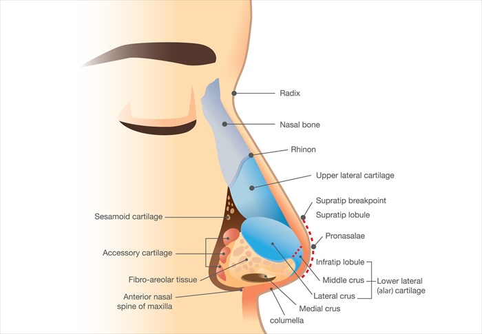
The nose can be divided into thirds, namely the lower, middle and upper regions. The lower third describes the nasal tip which consists of the lower lateral nasal cartilages, nasal septum and the nasal tip. The middle region or third is made up of the upper lateral cartilages and the nasal septum. And finally, the upper region or third consists of the bony skeleton at the top of the nose.
When it comes to non-surgical rhinoplasties, practitioners use a number of terms to describe the various nasal structures that may be treated. These are as follows:
- Radix – This refers to the root of the nose that correlates with the upper end of the nasal dorsum and is centred as the nasion. Simply put, this is the area of the nose that is formed where the forehead meets the nasal bridge.
- Nasal dorsum – This is also known as the nasal bridge and forms the majority of the nasal structure that connects the face to the tip of the nose (from between the eyes to the tip), forming a bridge-like structure. This area has a substantial influence over the overall aesthetic appearance of the nose. It consists of the uppermost two-thirds of the nose, including the middle cartilage as well as the upper bony areas.
- Nasion – The area refers to the depression easily seen at the nose’s root which corresponds with the nasofrontal suture (joining of the top of the nose to the face).
- Alar cartilage – This area offers support and structure to the lower portion of the nose. The alar cartilage consists of a cartilage pair, also known as the lower lateral cartilages. This cartilage forms a dome-like arch in the nose and consists of the columella and nasal tip.
- Rhinion – This is where the middle of the nose meets the top of the nose and refers to the area where the nasal bones (i.e. bony section of the nose) connect with the cartilage area of the nose.
- Nasal tip – This protruding area is the most forward and prominent part of one’s nose where the two lower lateral cartilages join together to form the tip. The nasal tip is divided into the infra-tip lobule, supra-tip and tip lobule.
There are a number of other slightly more complex terms that you may hear your medical practitioner use, however, the above list will allow for you to have a basic understanding of some of the most common ones and what they mean.
Common deformities of the nose
The below information outlines some of the most commonly seen irregularities and/or deformities of the nasal region:
**My Med Memo – A non-surgical rhinoplasty can correct these issues, however, their significance and what is required to achieve the best end result will be assessed by a doctor who will determine whether an NSR or surgical rhinoplasty is best suited to address the issue and achieve the desired results.
- Tip rotation – This refers to the position of the tip of the nose along an imaginary longitudinal rotation curve. Nasal tips are assessed according to their rotation, contour definition and projections. A tip that is over-rotated will point upwards towards one’s nose, this makes it appear more pointy or perky, some people may refer to this as a “Miss Piggy” nose. A tip that is under-rotated will point downwards making it appear longer and droopy.
- Saddle nose – This deformity refers an indentation in the nasal bridge as a result of cartilage inflammation which leaves a clear step and rather deep depression between the rhinion and bony upper part of the nose – thus resembling something of a saddle on the nose. A saddle nose is the result of damage to the supporting structures of the alar cartilages, leaving a depression or ‘dent’ that is saddle-like in appearance. The most common issues that lead to the development of saddle nose include trauma due to impact, autoimmune diseases (constant inflammation of the nose due to infection can result in the breakdown of supporting cartilage), cocaine abuse and overly aggressive surgery.
- Tension nose – This deformity of the dorsal area of the septum occurs due to overgrowth giving the nose a high, narrow and overly-projected appearance. A tension nose is characterised by excessive nasal protrusion, a downward tip and a defined dorsal hump on the nasal bridge.
- Polly beak – This is a complication that occurs post-surgery and refers to the typical appearance of the dorsal nasal curvature resembling that of a parrot’s beak.
- Retracted columella – The columella refers to the cartilage bridge and skin situated between the two nostrils. When looking at the nose laterally and the columella is not visible and protruding by two millimetres or more, it is then classified as a retracted columella. This may be the result of a birth defect, trauma or overly aggressive surgery.
- Hanging columella – This deformity refers to an overgrowth of the nasal septum which causes an overly-protruding columella.
- Pinched lobule – Also known as a pinched nose, this deformity is often the result of overly aggressive surgery. It occurs when the alar cartilages in the nose are either overly tightened or trimmed resulting in the domes within the alar cartilages collapsing, creating a pinched appearance.
- Bulbous tip – This deformity occurs when the alar cartilages are irregularly arched or broad, resulting in the development of what looks like a ‘ball’ at the tip of the nose.
- Inverted ‘V’ – This deformity occurs post-surgery and is the result of the upper cartilages in the nose detaching from the nasal bony areas, leading to the appearance of an upside down ‘V’ at the connecting area of the middle and upper area of the nose.


