Written by: Bronwen Watson
What other types of benign skin growths can occur?
- Dermatosis papulosa nigra (DPN): Similar in appearance to seborrheic keratosis or skin tags, these growths can occur commonly on the face and neck, as well as the back or chest. The growths are also more common among darker complexions. Growths are typically small, and black or brown in colour, occurring in isolated spots (a few or on many areas of the body). DPNs may be flat or as raised growths that hang off the body (similar to a skin tag). Causes are unknown, but these growths are not normally a cause for concern. A diagnosis can be made by sight, rarely needing a biopsy. Treatment can help to minimise the appearance of these growths, instead of providing complete removal. Affecting skin with darker pigment, a person is at risk of developing pigmentation defects (such as a lightening or darkening of the skin), as well as scarring (giving a blotchy appearance). Treatment can be administered by means of scissor or shave excision (removal), electrodessication, cryosurgery, dermabrasion or laser removal.
- Sebaceous hyperplasia: When the sebaceous glands surrounding a follicle in the skin enlarge, these growths occur. They are typically characterised as flesh-coloured or yellowish papules with a central dell (resembling a bump with a dimple or crater) and range from 2 to 9mm in size. These growths often occur on the facial area, especially the cheeks, forehead and nose, but can also occur on the mouth, chest (especially the areola – ring of pigmented skin around the nipple), or vulva (outer portion of the female genitals) and scrotum, penis shaft or foreskin (male genitals). It is thought that facial sebaceous hyperplasia is caused by declining androgen circulation due to aging. A biopsy may be warranted if the growth resembles a basal cell carcinoma (form of skin cancer). Laser therapy or surgical removal procedures can be performed to treat these growths.
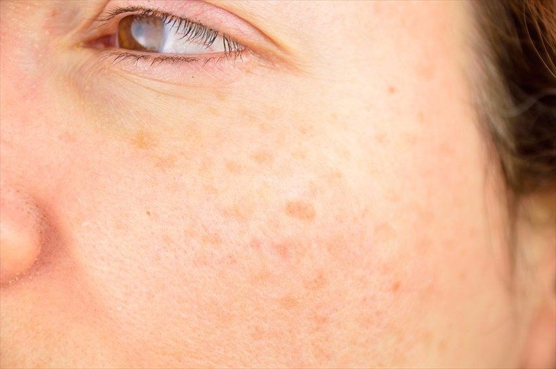
- Lentigines (liver spots): Often resembling moles (nevi), these hyperpigmented macules (patches) are usually a pale tan or brown colour. Lentigines commonly occur on sun-exposed skin areas, such as the face, neck, shoulders, back, chest, forearms and hands. These markings mostly indicate excessive photodamage which can result in an increased risk of sun-induced skin cancer. Rapid growth and changes in colour are typical indicators for a doctor to perform a biopsy and evaluate further. Treatment, however, is usually for cosmetic purposes and involves cryotherapy, laser therapy, bleaching creams or chemical peels.
- Cherry angiomas (senile angiomas): These growths are common in adulthood and are usually classified as ‘benign vascular growths’. These small, red circular or oval papules or macules (that appear cherry-like) can occur in most areas of the body, but seem to frequent the chest, abdomen or back areas more often. The growths are often smooth or even on the skin, but can also appear raised. These growths are typically asymptomatic, but can bleed when exposed to injury. Broken blood vessels in the growth give it a cherry-red appearance. Changes in size, shape or colour are usually indicators for concern (carrying a risk of skin cancer), especially if bleeding occurs. In this case, a biopsy may be necessary for assessment. Causes are linked to chemical exposure, climatic elements, as well as pregnancy. Treatment is, for the most part, unnecessary, unless removal is needed for medical reasons. Regular bleeding due to constant friction (bumping against the affected area) or cosmetic purposes are other reasons these growths may be removed. Treatment options include electrocauterisation (burning with an electric current), cryosurgery, laser surgery (using a pulsed dye laser), or shave excision.
- Epidermal inclusion cyst (EIC): These growths are typically caused by injury or trauma to the skin, such as following a minor surgery or as a result of body-piercings. A firm-to-the-touch cyst (fluid-containing membranous sac) forms at the site of injury when the surface of the skin (epidermis) gets into the deeper layers (dermis). Cysts may be flesh-coloured or reddish and inflamed (the skin covering the nodule), but are usually painless, and are not normally itchy. Singular growths are more common than multiples and can range in size. A physical examination can be done to diagnose the cyst. Ways in which a cyst may be physically assessed include a dermascopy (magnified lens), a Wood’s lamp examination (using ultraviolet light) or skin biopsy (although this is not usually necessary unless there is a need to rule out other potential medical conditions). Removal of the cyst can be done if bothersome (or if it ruptures which can cause pain) or for cosmetic reasons and usually involves a surgical excision (complete removal of all cystic contents). Treatment following removal may require warm compresses, corticosteroids or systemic antibiotics to prevent a secondary infection. If determined, an underlying injury or medical condition may also require treatment.
- Milium cyst: Often referred to a ‘baby acne’ (although inaccurate), a milium cyst is a small, white bump which often surfaces on the nose, eyelids and cheek areas of the face. Cysts can occur as groupings (milia) and form when keratin (protein) becomes trapped beneath the skin’s surface. These cysts are most common among new-borns (from the time of birth), but a cause is yet to be distinctively determined. These growths can occur in older children and adults too, usually as a result of damage to the skin, such as burns, blistering injuries, long-term sun damage or following skin resurfacing procedures (such as laser resurfacing or dermabrasion). Cysts are not normally accompanied by any inflammation or swelling. Diagnosis is made based on physical appearance (a physical exam), and very rarely requires treatment. Usually these cysts resolve themselves within a matter of weeks. If there is a degree of discomfort or pain, these cysts can be treated with chemical peels, deroofing (cyst contents are picked out using a sterile needle), topical retinoid creams, laser ablation, cryotherapy, diathermy (extreme heat) or destruction curettage (surgical scraping).
Find this interesting? Share it!

