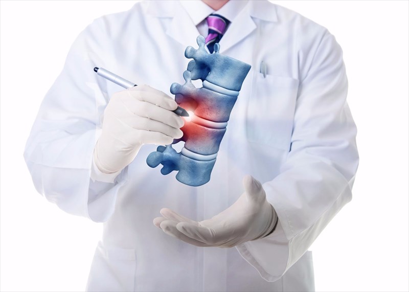
- Spinal stenosis
- What causes spinal stenosis?
- Location and types of spinal stenosis
- How does spinal stenosis affect the body?
- Who is at higher risk of developing spinal stenosis?
- How is spinal stenosis diagnosed?
- What is the treatment for spinal stenosis?
- Coping with spinal stenosis
- What is the outlook for someone with spinal stenosis?
What is the treatment for spinal stenosis?
Once a doctor has determined the location of stenosis and diagnosed a type, as well as the severity of compression on the spinal cord, appropriate treatment can be implemented.
1. Non-surgical treatments
Mild symptoms may be treated with non-surgical treatment options and regular follow-up appointments will be required to monitor a person’s overall condition. These include:
- Medications: Remedies for pain relief involve medications including NSAIDs (non-steroidal anti-inflammatory medication) such as aspirin, ibuprofen and naproxen, and other analgesia such as acetaminophen / paracetamol. Low doses of these over-the-counter medications can offer some short-term pain relief, and should be carefully monitored by a treating doctor. ‘Nerve block’ injections (anaesthetic) can also be used for pain relief. Muscle spasms and degenerated (damaged) nerves may also be temporarily relieved with anti-seizure medications and muscle relaxants. Inflammation in the neck and back can be alleviated with corticosteroid / epidural injections (a commonly used medication is prednisone), directly targeting the source of symptoms. Injections (in larger doses than oral versions) are normally used sparingly due to the nature of side-effects (i.e. repeated injections can weaken connective tissue and other nearby bone structures), but can help to alleviate swelling and irritation directly at the nerve roots. Injections do not prevent stenosis, but can relieve some degree of pain by alleviating inflammation. Morphine-related medications (opioids), such as oxycodone or hydrocodone may provide some short-term relief of symptoms as well. These will be managed and monitored carefully on a prescription basis as they do carry side-effect risks and can become habit forming.
- Assisted devices: To aid in easing movement, especially walking, corsets, walkers and braces can be used.
- Exercise: Balance, flexibility and strength can be improved somewhat with regular physical activity. Symptoms of stenosis can actually be better treated with activity than adopting a less active or sedentary lifestyle. Muscles which weaken as a result of reduced activity can in fact lead to more symptoms of pain in the body. It is advisable to consult a physical therapist for advice and guidance when it comes to activity types which are best suited for the maximum benefit. For instance, those with lumbar spinal stenosis who are more comfortable in forward flexed positions may benefit from stationary biking (leaning forward on handlebars) instead of trying to walk for exercise. Physical therapy, while not a cure, can help to build up strength and endurance levels, as well as improve stability and flexibility in the spine, which helps with balance too. Physical therapy can help to get a person going in the right direction, building strength and tolerance slowly. Once comfortable, a person can transition into their own regular exercise routine and continue to enjoy the benefits of activity.
- Percutaneous image-guided lumbar decompression (PILD): A decompression procedure, a doctor can use needle-like instruments to remove a portion of a thickened ligament in the spinal column in an effort to create more space in the spinal canal, and alleviate a spinal root impingement. This procedure is commonly done for those suffering from lumbar spinal stenosis with the development of thickened ligaments. The procedure is performed without the need for general anaesthesia and is thus classified as minimally invasive (non-surgical). If a person is high risk for a surgical procedure, a doctor may consider PILD as a treatment option as it carries fewer risks.
2. Surgical treatments
Where stenosis has resulted in severe symptoms or has not responded well to non-surgical treatments and advised homecare tips, surgery is the next best option. Due to the degenerative nature of spinal stenosis, symptoms can worsen, becoming disabling (due to permanent damage).
Surgery can help to relieve pressure on the spinal cord and spinal nerve roots, by creating more space in the spinal canal. Decompressing (using surgical decompressive techniques) the area where stenosis occurs has proven the most effective way to treat, and resolve, symptoms of the condition.
The diagnostic process is essential before any surgical procedure can be considered. Imaging tests, in particular, provide the most comprehensive means to determine a positive surgical outcome by identifying all areas of compression along the spinal canal. If this is not established, the chances of achieving symptom relief will be somewhat inferior.
Today’s modern imaging techniques allow for fewer operative ‘surprises’ (spinal instabilities once a patient has been opened up), which can also be detected and even anticipated (and planned for) ahead of time.
Surgical technique options include:
- Laminectomy (or decompression surgery): The back portion of a vertebra is known as the lamina. During a laminectomy, a surgeon will remove this portion of an affected vertebra, easing pressure on the spinal nerves and creating the space necessary to alleviate symptoms caused by narrowing and inflammation. The vertebra can sometimes be linked to an adjoining vertebra with the use of metal hardware (a metal plate) and a bone graft (known as a spinal fusion technique) to help reinforce the spine (creating more strength in the spine). This is necessary if too much bone needs to be removed, or if there is excessive motion between the bones and more stability is required. This surgery is most often used for lumbar spinal stenosis, but can also be performed for cervical spinal stenosis (often combined with a fusion).
- Laminotomy: Only a portion of the lamina is removed during this procedure (not the entire back portion of the affected vertebra). A hole is carved into the affected lamina, which effectively helps to relieve pressure within a particular spot.
- Laminoplasty: A surgeon may consider this technique for cervical vertebrae, opening up the space within the spinal canal by using metal hardware bridges – much like a hinge which creates more space while bridging the gap within an open portion of the spine. This procedure is often used for cervical spinal stenosis.
- Anterior cervical discectomy and fusion (ACDF): This procedure is one of the most common treatment procedures for cervical spinal stenosis. A surgeon will remove a disc between two vertebrae, as well as any other abnormalities, like bone spurs which may be placing pressure on the spinal cord nerves. A removed disc will be replaced with a bone graft and metal hardware so as to fuse bone together (so that it can grow together).
- Corpectomy: A procedure wherein one or more vertebrae are removed and multiple fusion techniques are used.
Other options available may include:
- Interspinous process spacer: Spinous processes are small prominences which run along the back of the spinal column, near the skin’s surface. As these are in near proximity to the skin, interspinous process spacers can be implanted with minimal intervention, invasiveness and associated side-effects or complications. The devices open the foramen (where spinal nerve endings travel away from the centre of the spinal canal) and limit spinal extension (i.e. the position of the spine when bending backward which is very painful for those with stenosis). Devices are implanted while the patient is under mild sedation (local anaesthetic) as an outpatient procedure (requiring no overnight stay in hospital) and may be beneficial for those who are high risk for more extensive surgical procedures such as the elderly.
- Foraminotomy: Also a decompressive surgical technique, the area where the spinal nerve root exits the spinal canal is made larger (foramen), creating a passageway. Bone or tissue which is obstructing this passageway, compressing the spinal nerve root, is removed.
- Microendoscopic decompression / microendoscopic laminectomy (MEL): An alternative to a traditional laminectomy, a MEL procedure offers a less invasive approach to achieve the same decompression goals. A state-of-the-art endoscope is used for visualisation purposes, while a thin needle is placed under fluoroscopic guidance (X-ray). A surgeon will make a small incision around the needle, which is then guided to the affected bone. A hollow, metal cylinder is then passed to where stenosis is occurring and secured in place. A rigid surgical microendoscopic camera is placed inside the cylinder to allow for magnified views of the area being operated on. A surgeon then micro-surgically removes the bone causing stenosis, alleviating pressure on the spinal nerve roots. This procedure is also effective for removing ligamentum flavum and herniated discs and reduces trauma or damage to soft tissues, which also enables a faster recovery period following surgery.
A patient is required to stay in hospital for at least 1 to 2 days following a surgical procedure. If a fusion has taken place during surgery, a brace may need to be worn during the initial days of recovery.
Following surgery, pain and inflammation is normally managed with prescribed medications for a set period of time (normally between 4 to 8 weeks). Medications can be habit forming and thus will only be prescribed for a limited period, and thereafter over-the-counter drugs may be used if necessary.
Those who have a spinal fusion may not be able to use NSAID medications for at least 6 months following surgery as these can cause bleeding and interfere with the healing of affected bones.
A surgeon may also recommend the following care instructions following surgery:
- Seek medical attention immediately if signs of infection arise – a high fever / temperature, drainage, pain, redness and swelling
- Refrain from smoking as this delays healing and increases a person’s risk of other health complications.
- Wear a provided brace as instructed by the treating surgeon.
- Refrain from driving at least 2 to 4 weeks after surgery.
- Request help from a loved one with certain daily activities such as dressing or bathing during the initial few weeks of recovery. A shower may be permitted only on the 4th day following surgery (unless instructed otherwise)
- Avoid being in a sitting position for long periods of time (try and get up and move around carefully every so often).
- Avoid lifting heavy objects, and do not bend or twist from the waist.
- Refrain from doing any housework (i.e. vacuuming, ironing or loading and unloading a washing machine or dishwasher) or gardening until deemed safe by the treating surgeon.
- Do not engage in sexual activity until deemed safe by a treating surgeon.
- Gradually return to normal activity (i.e. start with short walks and physical therapy)
Surgical procedures do carry risks and can result in damage to nearby healthy tissues, post-surgical pain or inflammation, infection, internal bleeding, anaesthetic reactions, tears within the membrane of the spinal cord (or dural tears), cerebrospinal fluid leaks, blood clots and neurological deterioration.
Other complications may involve vertebrae that fail to fuse (often as a result of existing problems such as smoking habits / nicotine which inhibits bone-growing cells, obesity, osteoporosis or malnutrition), deep vein thrombosis (DVT), bone graft migration (the graft moves from the placed position following surgery), a hardware fracture (screws, plates and rods used to secure and stabilise the spine move or break before fusion takes place sufficiently), bone graft migration, or transitional syndrome (vertebrae above or below the treated portions of the spine take on additional strain or stress).
Before any surgery takes places, risks and benefits will be discussed in full, having been comprehensively assessed by the treating doctor ahead of being recommended.
Whether a person is undergoing non-surgical treatment or receives a surgical procedure to alleviate symptoms of stenosis, periodic / regular follow-ups with a treating doctor will be recommended. If mild symptoms occur, follow-ups may be frequent or as and when required (i.e. when symptoms begin to worsen or new ones develop). Follow-ups post-surgery may continue for years afterwards, depending on the nature of a person’s condition.
What kind of research is being done to improve treatment for stenosis?
To date, the most effective way to alleviate symptoms of stenosis is to create space within the spinal canal using decompression techniques.
Researchers have been testing the use of stem cells in an effort to come up with a regenerative means to treat this degenerative condition.
Genomics (molecular biology and genetic material studies) are another means being clinically trialled to determine potential gene therapies which can help to resolve stenosis.
