- Spinal stenosis
- What causes spinal stenosis?
- Location and types of spinal stenosis
- How does spinal stenosis affect the body?
- Who is at higher risk of developing spinal stenosis?
- How is spinal stenosis diagnosed?
- What is the treatment for spinal stenosis?
- Coping with spinal stenosis
- What is the outlook for someone with spinal stenosis?
What is spinal stenosis?
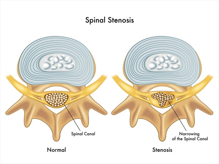
Spinal stenosis is a medical condition wherein the spinal column compresses the spinal cord as a result of the narrowing of the spinal canal. The spine, a key portion of the central nervous system, connecting the brain with the rest of the body, is essentially a column of connected bones (stack of bones known as vertebrae) that provide stability and support for the upper body (much like ‘shock-absorbing’ discs). It is this bodily structure which enables us the ability to move, turn and twist.
Nerves run through the vertebral openings and channel signals from the brain to the remainder of the body. Tissues and bone surrounding these nerves offer some protection, allowing for ease of movement, balance and sensation. An impairment which affects these tissues can interfere with these functions, causing pain and discomfort.
Part of the ageing process, spinal stenosis typically happens gradually with symptoms only really becoming apparent when narrowing within the vertebral openings pinch (tighten) and place pressure on the nerves. When this becomes excessive, it effectively compresses the vertebral nerves which travel through the length of the spine. Narrowing or compression can occur at any point along the length of the spine but are most commonly experienced in the neck and lower back. When nerve compression occurs symptoms of tingling and numbness (often in the torso, arms and legs), muscle weakness, as well as pain may be experienced. Such symptoms can worsen over time as more compression occurs.
To date, there is no cure for spinal stenosis, but there are variety of treatment options available for the effective management of symptoms, without compromising quality of life too greatly.
Spinal stenosis and the structure of the spine
Stenosis, meaning ‘the abnormal narrowing of a bodily structure or channel’ (and comes from the Greek word which means ‘choking’), results in the compressing or squeezing of, in this instance, the spinal cord and its nerves.
The upper portion of the spinal canal (the cavity which runs from the head, through each of the vertebrae to the pelvis, enclosing the spinal cord) is known as the cervical spine (the neck). The middle portion of the spinal canal is known as the thoracic spine (in the mid back area). The lower portion of spinal canal is known as the lumbar spine (in the lower back).
The spinal cord effectively travels from the brain, through the spinal canal, down the back, and ends at the pelvis (the bottom of the spine is known as the sacrum). All along this stack or column of bony blocks, which support the body, are additional bony attachments. These help to stabilise the spine, as well as protect the spinal cord and nerves along the length of it between the brain and other nearby organs, muscles and sensory structures. All bony attachments and discs are referred to as spinal segments.
Spinal nerves and the spinal cord in the spinal canal, the fluid-filled sac which acts much like a protective garden hose casing, are moveable and can bend along with the spine. The outer membrane of the sac is known as the dura mater (tough mother).
Nerves which exit the spinal cord at each vertebral opening (holes or nerve roots known as neuro-foramina) between the neck and the lower back are responsible for controlling the movement of the body’s limbs (arms and legs) and sensation of the skin. Nerves extending off of the spinal cord at the base of the spine are known as cauda equina (the Latin name for a horse’s tail, which they resemble).
Stenosis typically occurs as a result of degenerative changes (or bony growths) which constrict the normal spaces between the spinal cord or nerve roots.
Other Articles of Interest

Scoliosis
Scoliosis is a condition that refers to a rounding or curvature of the spine and is commonly seen in children. Find out more about the condition here...

Bunions (Hallux valgus)
Bunions, also known as hallux valgus, is a progressive disorder affecting the big toe at the base of the joint. An inward leaning and rotation of the big toe characterises this condition. Learn more.

CT (Computerised Tomography) Scan
What is a CT scan and what exactly does it involve? We break down this testing procedure so that you know what to expect when you go through your scan.


