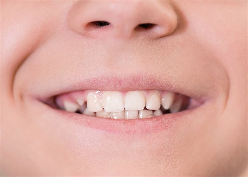
- Teething
- Dental anatomy
- Primary tooth eruption
- Stages of teething
- The most obvious signs that your child may be teething
- Signs and symptoms that are not associated with teething
- Tips to help soothe a teething child
- Things to avoid when trying to soothe a teething child
- How to care for your child's teeth and gums
- When should you take your child to the dentist for the first time?
Not only do they play a pivotal role in how we speak and contribute to our overall appearance, but teeth essentially determine how we chew and our incising, shredding, and crushing potential, which is important for digestion.
Each individual tooth has an unmistakable crown that is located above the gum with often more than one root ranging into the alveolar bone (this is the condensed rim of bone that essentially keeps teeth in place) of the maxilla (upper jawbone) or mandible (lower jawbone). A peg and socket seam is formed by the tooth within the alveolar bone and the tooth is kept in place by the periodontal membrane that permits slight movement thereof.
Teeth are comprised of four layers:
- Enamel
- Dentin
- Pulp
- Cementum
Enamel
Covering the entire surface of the crown of each tooth, safeguarding it against decay, cracking and wear and tear, dental enamel is the body’s hardest tissue. It is comprised of 96% mineral content, 2% water, 1% protein, and 1% other components. This structure makes the tooth strong and resistant, forming a barrier against acid and plaque, as well as heat and cold, protecting the inner layers of the teeth.
Once enamel is destroyed, it cannot be regenerated by the body, as such, it is imperative that it is protected through proper dental hygiene practices, and by avoiding foods that may damage it. While primary teeth eventually fall out and are replaced by permanent teeth, it is still important to maintain the condition of your child’s teeth in order to prevent pain and compromised nutritional intake as a result.
Dentin
Dentin is comprised of approximately 60% mineral and 20% organic based compounds including protein (collagen type 1). It lies just below the enamel, acting as a support structure for it and forming the bulk of tissue of the tooth. It ultimately determines the shape and size of the teeth.
Dentin is produced by the dental pulp as a form of protection, and the production thereof is elevated in response to environmental factors such as tooth decay and damage. When this occurs, dentin is delivered to the pulpal wall to make any necessary repairs and defend the pulp against further damage.
Pulp
Dental pulp is a soft, gelatinous, highly specialised living tissue situated at the centre of the tooth, consisting of odontoblasts (cells that produce dentin), blood vessels, fibroblasts (cells that synthesize collagen), nerves, and a complicated extracellular matrix. It extends throughout the length of the tooth and contributes to the neurosensory activity and repair function of the teeth.
Cultivating a healthy dental pulp until the root of the tooth is completely developed and has walls broad enough to bear the brunt of the force generated by the crown during biting and chewing is essential.
Cementum
Cementum is the layer of tissue (made up of collagens and proteins) that has been mineralised and covers the tooth’s root which is located inside the socket of the gum itself. The tooth is kept in place inside the jaw by 4 periodontal elements which include the following:
- The gums
- The jawbone
- Cementum (which only envelopes the tooth’s root)
- The periodontal ligament
Cementum is comparable to bone with the exception that there is no blood or nerve supply to it. Adding to this, cementum will only move when the tooth itself moves.
How does cementum work?
The core aim of cementum is the attachment of teeth to the collagen fibres within the periodontal ligament. This will help to maintain the roots of the teeth as well as their position inside the gum and the bone itself. Cementum also plays a pivotal role in repairing and regenerating teeth.
Once a person ages, their gums will eventually start to recede (i.e. pull back from their original position) and the roots of the teeth may become visible. This can result in the cementum being eroded and dentin becoming exposed.
