- Raynaud Phenomenon / Syndrome / Disease
- What causes Raynaud phenomenon?
- What are the signs you may have Raynaud phenomenon?
- Risk factors for Raynaud phenomenon and potential complications
- Diagnosing Raynaud phenomenon
- Treating Raynaud phenomenon
- Living with Raynaud phenomenon
- Raynaud phenomenon FAQs
Which doctor to see if you think you may have Raynaud's syndrome
- General practitioners (GPs or family physicians)
- Internists
- Rheumatologists
- Hand surgeons / specialists (often with certified training in other specialised areas, such as orthopaedic surgery, plastic surgery, or general surgery)
What to expect during a consultation for Raynaud's disease
A consultation with a medical doctor will usually begin with a discussion regarding symptoms. Generally, a doctor will ask a series of questions in order to obtain as much detail and clarity about symptoms as possible.
Some questions which are likely to come up may include:
- How long ago did symptoms begin?
- How frequently do symptoms (i.e. ‘attacks’) occur?
- Have any colour or sensation changes been noted in the affected areas? (Such as numbness or pain in the toes and fingers, or colour changes ranging from white, blue and red / flushed)
- Does anything specific appear to trigger symptoms?
- Is there a history of rheumatic illnesses in the family?
- Is there any member of the family with similar symptoms or a diagnosed Raynaud's condition?
- Are any other medical conditions known to you? (I.e. have you been diagnosed with any other illness?)
- What medications or supplements have recently been taken (or are currently being taken)?
A doctor will also wish to gain some clarity on certain known triggers or symptom aggravators, such as caffeine use and smoking habits. Occupations or regular activities (i.e. recreational) may also be questioned to determine any potential repetitive movements which may be considered triggers or aggravators for symptoms of Raynaud phenomenon.
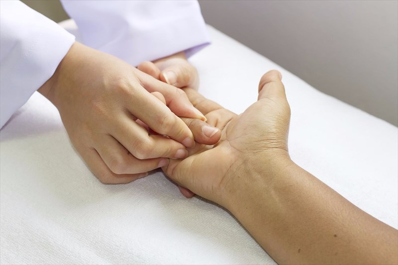
Once enough information is obtained to use as a base for assessment, a doctor will conduct a physical examination of the affected areas of the body.
Testing for Raynaud phenomenon
There is no single diagnostic test for Raynaud phenomenon, but a doctor may recommend one or more so as to rule out any other underlying conditions which may be related to symptoms of the illness. In this way, testing may be used to make a diagnosis and determine the type of Raynaud phenomenon present (i.e. primary or secondary). Testing may also largely depend on the symptoms present in the patient.
Tests can involve:
A cold stimulation test: This test may be used as a way to trigger a vasospasm attack. A doctor may present a small temperature measuring device which will be attached to fingers or toes. Hands or feet will then be placed in ice water for several seconds and then removed. The exposure to ice cold conditions aims to trigger symptoms and then assess how long it takes for the skin to resume a normal body temperature using the measuring device. Although uncomfortable, the test poses no health risk to a patient and can be done immediately during a consultation. A doctor will assess discolouration changes, and discuss any sensations experienced as well. Temperature which returns to normal within 15 minutes is usually considered (reasonably) normal. Test results may be used in conjunction with any or all of the below evaluations.
- Capillaroscopy: This is a microscopic examination of the nailfolds (fingernails or toenails) and is commonly used to determine differences between primary and secondary Raynaud phenomenon. Those with the secondary form of the condition typically have enlarged blood vessels or capillaries near the nailfolds (deformities can also be detected). Capillaries are typically ‘normal’ in those with primary Raynaud phenomenon, when a vasospasm attack is not active. The test is often performed in the doctor’s office and could be used as a starting point for further testing should a doctor feel it necessary.
- Antinuclear antibodies (blood test): This blood test will help to determine primary from secondary Raynaud phenomenon conditions by assessing the presence of ANAs (antinuclear antibodies), produced by the immune system, in the bloodstream. If these proteins are present (a sign of a stimulated immune system), it may be one indicative marker of an autoimmune or connective tissue condition.
- Erythrocyte sedimentation rate (ESR or sed rate test): Another blood sample can be analysed (in a laboratory) to determine the rate at which red blood cells settle into the bottom of a test tube. If this process of descending cells is determined as faster than normal (i.e. the standard rate), it could indicate a potential underlying rheumatic / inflammatory or autoimmune disorder (i.e. an elevated sed rate due to inflammation in the body).
- C-reactive protein blood test: CRP or C-reactive proteins are normally produced by the liver in response to inflammation in the body. A high-level present in the bloodstream may indicate inflammation in the arteries connecting to the heart or those associated with any other inflammatory condition, such as rheumatoid arthritis or lupus.
- Rheumatoid factor blood test: High levels of proteins produced by the immune system in the blood could indicate inflammatory conditions.
If testing does not show up any diagnostic markers for other associated conditions, it is likely that a doctor will diagnose primary Raynaud phenomenon. If testing does present inflammation markers, it is likely that a doctor will look for (using a physical exam and appropriate testing) signs of other conditions. For instance, thickened skin could indicate scleroderma or a malar rash (‘butterfly shape’) characteristic of lupus.
Symptoms of Raynaud's can develop years before an inflammatory condition. Thus, a doctor could diagnose primary Raynaud phenomenon, but request periodic check-ups to monitor any possible condition developments. If any connective tissue disorders are suspected, it is likely that a GP or internist may recommend a Rheumatologist visit as these specialists typically have more knowledge and experience with these associated conditions.

 A cold stimulation test: