If an ocular migraine is suspected, it is best to consult a primary healthcare professional (family physician or general practitioner / GP) or an optometrist.
There isn’t a specific set of tests which will diagnose an ocular migraine condition, but a comprehensive medical evaluation is necessary in order to either determine or rule out potentially serious underlying causes, as well as to effectively treat a person’s condition.
The first thing a doctor will wish to establish is the nature of symptoms being experienced, and note a full medical history (including a family history of medical conditions, especially if similar headache characteristics are evident). To do this a doctor will ask a series of specific questions.
How clear a patient is regarding their symptoms and medical history has a considerable influence, helping a doctor to make the most accurate diagnosis. It can be helpful to prepare a few notes before a consultation which detail notable changes before the attack, as well as what is experienced during and after it. It is helpful for a physician to know where any sensation changes or pain occurred (the exact location), how long the attack lasted (its duration), and what symptoms other than visual changes and pain were experienced.
A physical examination may be performed to assess a person’s overall health condition and check things such as blood pressure and other vital functions (heart rate and breathing). A physical exam won’t necessarily help a doctor pinpoint an exact cause of the migraine experienced prior to the consultation but it is helpful to assess the patient’s current health status. A diagnosis may need to be made following a process of exclusion (ruling out potential underlying conditions) and in this way, determine ocular migraine as a possible explanation for transient visual disturbances.
Some conditions or problems which can cause a similar set of symptoms include:
- Amaurosis fugax (a temporary, but painless loss of vision affecting the eyes due to lack of blood flow to the retina)
- Giant cell arteritis (inflammation of the blood vessels of the scalp)
- Arterial spasms
- Drug abuse
- Polycythaemia (a high concentration of red blood cells)
- Sickle cell disease
- Stroke
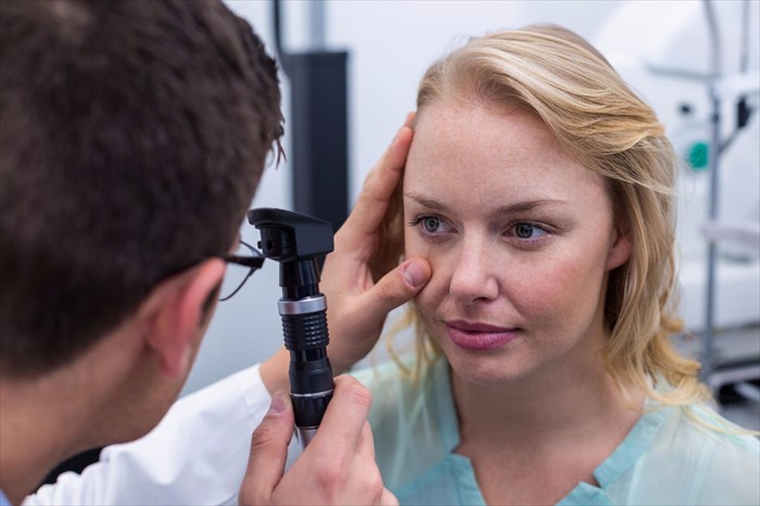
During the consultation, a doctor may make use of an ophthalmoscope (also known as fundoscopy), which is a screening tool that has a light attachment and numerous small lenses to check for abnormalities or damage at the back of the eye (the fundus – retina, optic disc and blood vessels). This screening test may be used to assess a potential reduction in blood flow, or damage caused as a result, to the affected eye. It isn’t likely that a diagnosis will be made using this test alone. Restricted blood flow can occur as a result of various other conditions including hypertension or diabetes, and generally takes place during a migraine attack (while blood vessels are narrowed). It may be best to perform a fundoscopy during an ocular migraine as a result. The doctor will make the call based on the nature of a person’s condition during the consultation, taking how long ago the attack was experienced into consideration.
If an eye screening test is to be performed, a doctor may choose one of three ways to perform it:
- Direct examination: A doctor will request a patient to sit in a chair before switching off the lights in the consulting room. The doctor will sit across from the patient and look through the lenses of the ophthalmoscope to examine the eye (using the light of the scope). A patient may be asked to look in various directions during the examination (up, down or to the side etc.).
- Indirect examination: A patient may be asked to lay down or be seated in a reclined position. This allows a doctor to see structures at the back of the eye in a little more detail. Using the bright light shining down on the affected eye, the lens of the scope will be held directly in front of the organ for examination. Again, a doctor may request some movement and ask that a patient look in various directions while assessing the organ. Sometimes a small, blunt probe may be used to apply a little pressure to the eye.
- Slit lamp examination: The view of the back of the eye using this technique is much the same as that which is achieved during an indirect examination. A slit lamp, however, provides greater magnification. A patient may have drops administered to dilate the pupils and will be seated and the slit lamp positioned across from him/her. He/she will be asked to keep their head steady by resting their chin and forehead on the instrument. A doctor will then shine a bright light directly into the eye and use a microscope to evaluate the back of the eye. Looking in different directions may be requested. Some pressure may be applied using a small probe.
A fundoscopy (with an ophthalmoscope) is not a comfortable screening test for a patient, but is not painful either. A temporary period of ‘afterimages’ (a visual that lingers in the field of vision following exposure to an original) may occur once the bright light has been turned off. Blinking several times should restore vision to the state it was in before the screening. If pupils are dilated, the wearing of dark glasses is advisable to avoid light sensitivity until they return to normal after a few hours.
Should any symptoms appear unusual, complex or quite severe, a doctor may recommend various other tests for either diagnostic purposes or a process of elimination. These can include a CT scan (computerised tomography) or MRI scan (magnetic resonance imaging). Both imaging tests can produce highly detailed, cross-sectional visuals of the inside of the body, showing functionality and abnormalities all over the body, including the head. An MRI makes use of a very powerful magnetic field and radio waves and can detect numerous abnormalities involving infections, tumours, blockages, bleeding and other characteristic indicators of neurological problems. A CT scan uses a series of X-rays to generate cross-sectional visuals and has, to date, helped to successfully determine the cause of many forms of headache.
Other testing options include blood samples which can be analysed in a laboratory. A doctor may wish to check for any signs of blood vessel abnormalities, possible toxins in the bloodstream or infections which may be affecting the brain and or/ spinal cord.
If there are signs of bleeding or infection, a doctor may recommend a spinal tap (also known as a lumbar puncture). This procedure involves the use of a thin needle which is pierced through the skin to reach between two vertebrae (bone structures making up the spine). A sample of cerebrospinal fluid is then extracted for laboratory analysis and any signs of bleeding or infection can be identified, assisting in the diagnosis of a possible underlying cause.
A doctor will take all evaluations made during the consultation and tests performed into consideration before making a diagnosis of ocular migraine. Characteristic symptoms, noted potential triggers and a ruling out of other serious conditions may also result in a diagnosis of ocular migraine.

