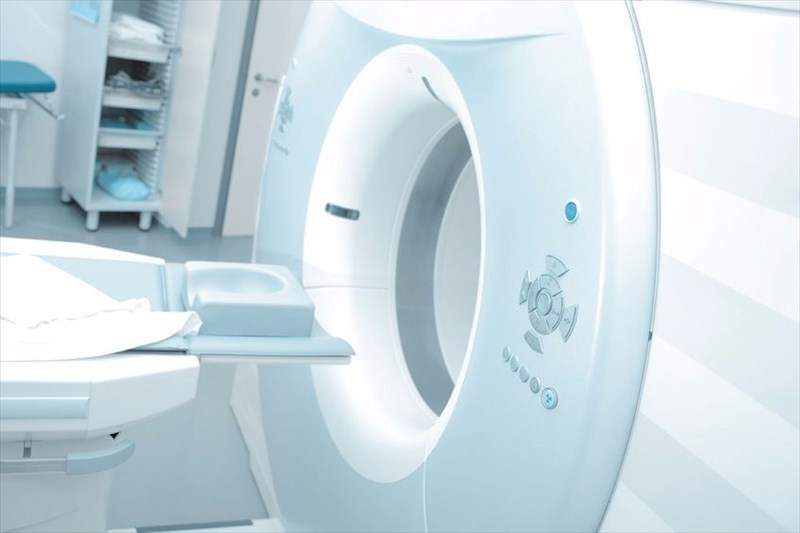
What is an MRI? (overview)
An MRI (magnetic resonance imaging) is a test that makes use of a magnetic field, as well as pulses of radio wave energy to produce pictures of organs, tissues and other structures inside the body.
This testing method can also give different information about the structures in the body that can also be picked up with an ultrasound, X-ray or CT (computed tomography) scan. An MRI can pick up other abnormalities or problems that cannot be seen with other types of imaging methods.
An MRI test allows for an area of the body to be studied inside a special machine that contains a strong magnet. MRI machines are large, tube-shaped magnets which allow for a person to lie inside. A magnetic field temporarily realigns hydrogen atoms in the body. Radio waves then cause the aligned atoms to produce very faint signals. These are used to create cross-sectional digital images (sometimes in 3-D or 3 dimensional visuals which can be viewed from many different angles), much like slices in a loaf of bread.
If available, an open MRI machine that doesn’t enclose the entire body may be used (these are not available everywhere and the image quality may not be as good as those from a standard MRI machine). Contrast material (or fluid) containing gadolinium can also be used during the scan to show up certain structures or parts of the body more clearly.
Digital images are created from an MRI scan and saved or stored on a computer to study later on or to be reviewed remotely.
An MRI is a non-invasive way for a medical professional to examine the goings on inside the body (organs, tissues, skeletal system and other structures). The high-resolution images that an MRI scan produces can help a doctor or specialist to accurately diagnose variety of problems or conditions.
Other Articles of Interest

Spinal stenosis
What is spinal stenosis? We take a look at the structure of the spine and how abnormal narrowing and constriction affects the ability to move, turn and twist...

Stroke
We all know that a person having a stroke is in a serious and life-threatening situation, but do we really know what to do in the event of an emergency?

Alzheimer's Disease
Do you find you or a loved one is suffering from progressive memory and mental deterioration loss in old age? It could be from Alzheimer's Disease...
