- Blood clot
- Types and causes of blood clots
- What risk factors contribute to blood clots?
- What are the signs and symptoms of blood clots?
- What kinds of blood clot complications can occur?
- How are blood clots diagnosed?
- What treatment procedures are involved in dealing with blood clots?
- Are there ways to prevent blood clots and what are the associated complications?
- Blood clot FAQs
Which medical professionals should you see if you think you have a blood clot
The type and location of the blood clot generally determines the best team of medical professionals required for optimum care (specialist care).
Medical professionals that may be sought should a clot be suspected include:
- Family physicians (general practitioners / GP)
- Internists
- Emergency physicians and critical care specialists
- Surgeons
- Interventional radiologists
- Cardiologists
- Cardiothoracic surgeons
- Neurologists
The doctor's visit
It’s important to consult a medical professional as soon as a suspicion of a clot becomes apparent. Any symptoms which arise suddenly must be attended to immediately, especially those concerning shortness of breath or breathing difficulty, light-headedness (low blood pressure), a cough that is accompanied by bloody sputum (phlegm), sudden changes in vision and chest pain or pressure.
If a person suspects a blood clot, it is a good idea to bring along a trusted family member to the medical consultation. If a complication arises or symptoms are severe, it may be very helpful to the attending medical professional to have someone around who can assist with medical history information, should the person experiencing a clot not be able to communicate themselves.
A doctor will begin a consultation with a discussion about the symptoms or concerns being experienced and obtain a detailed medical history. This is necessary so as to understand the nature of discomfort and also to determine potential risk factors.
He or she will be able to narrow down a potential location of a suspected clot during this discussion. Arterial blood clots seemingly develop suddenly (i.e. an acute event) and often cause serious symptoms very quickly. The body’s tissues are quickly deprived of oxygen (as the clot slows or stops the flow of oxygenated blood) which means that the onset of symptoms typically happens rapidly.
Sometimes, acute symptoms are accompanied by severe complications and signs of severe blockage, such as in the case of a heart attack. Symptom onset for venous blood clots happens a little more gradually, often progressing within a few hours of the clot development.
If a blood clot is suspected, a doctor will clearly explain essential information so that the person concerned, and their accompanying family member can understand the nature of the condition. It's important to understand that the location of the blood clot and the resulting adverse effects (restricted blood flow etc.) are of primary concern.
A physical examination will be conducted as a means to assist in determining additional information regarding a person’s physical condition. A doctor will assess a person’s vital signs and measure blood pressure, respiratory rate, pulse rate, body temperature and also assess oxygen saturation levels. He or she will use these measurements to assess how stable a person is in their current state. To assess a person’s heart rate and rhythm, a doctor may request an electrocardiogram (ECG / EKG). Any abnormal heart rhythms will be picked up on this electrical activity test procedure. If there are any signs of respiratory distress, a doctor may suspect a pulmonary embolus and listen for any abnormal sounds which may indicate damaged or inflamed lung tissue.
A doctor will look for physical signs which indicate clues as to the potential location of a clot to determine whether it is a venous or arterial thrombus.
- Venous blood clot: The affected area, such as a leg or arm, will likely appear red, swollen and be tender to the touch or painful.
- Arterial blood clot: The affected area may appear blanched (pale) due to a lack of oxygenated blood supply and feel cool or cold to the touch. A person may also feel numb (i.e. experience a loss of sensation) and unable to move (similar to a state of paralysis). Severe pain is another indication of an arterial thrombus.
Testing procedures for blood clots
A doctor will wish to try and determine the possible location of a clot in order to recommend appropriate testing. This helps to determine an accurate diagnosis and appropriate course of action for treatment.
The most effective tests to screen for a venous blood clot include:
- D-dimer (blood test): A small protein fragment known as FDP (fibrin degradation product), D-dimer is effectively the breakdown product of a blood clot. Elevated levels of the protein will be in the bloodstream and may indicate the presence of a clot, but this test is not used to make a definitive diagnosis. Rather, a doctor will pair results of this blood test (used as more of a screening test) alongside an imaging test in order to make a confirmed diagnosis of a blood clot. Elevated levels of D-dimer can also be seen in those with cancer or women who are pregnant.
- Ultrasound scan: One of the most common ways a doctor will make a diagnosis for blood clots is via an ultrasound scan. A doctor will apply a gel over the affected area and use a wand-like device known as a transducer to pick up any sound waves produced by a blood clot. Sound waves will be picked up from the body’s tissues and relay echoes back to a monitor, where a moving image will be reflected. A clot, if present, will then become a visible image on the monitor. A doctor can then determine the presence of a clot, its exact location and size. He or she will also be able to determine if the clot is immobile or on the move.
- Venography: An alternative test may be opted for if an ultrasound scan does not produce a clear result (although an ultrasound is most often preferred). A radioactive contrast dye will be injected into a vein in order to ‘show up’ a visual of an abnormality, such as a clot. The dye effectively ‘fills up’ the veins, providing a clear visual of the inside of blood vessels (veins). A doctor will then perform an X-ray (video X-ray) of the affected area. The visual result will help a doctor to make a diagnosis. A doctor will be able to determine the location of the clot and also see decreased flow of blood in the blood vessels.
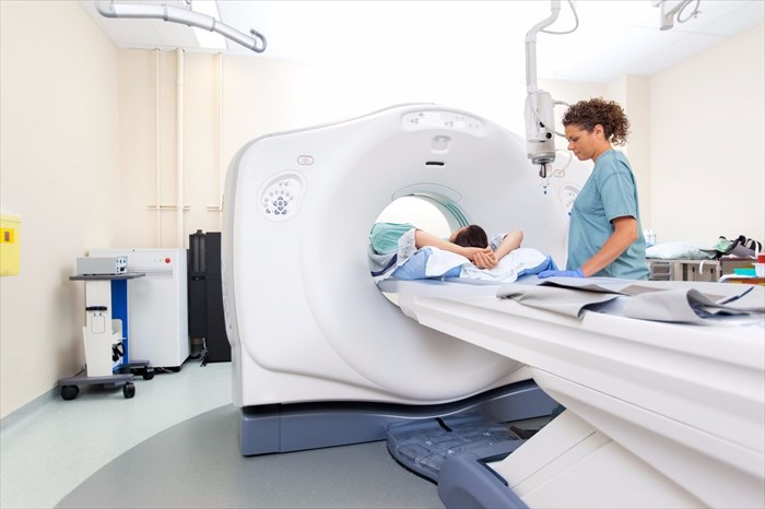
- Computerised tomography (CT) scan: In more severe cases, or if a doctor suspects a pulmonary embolus, a CT scan may be preferred over an ultrasound or venography imaging test. (9) A radiologist will be able to obtain clear and detailed images of the affected area of the body, and quickly pick up the presence of a clot, its location and level of concern for potentially serious complications. A contrast material may be injected intravenously to better show the pulmonary vessels on the scan and pick up a potentially mobile thrombus (embolus), as well as any damage that has occurred to vital organs such as the heart and lungs. A radiologist will be able to see whether the heart is straining to pump or whether the embolus is already present in a lung.
- Ventilation perfusion (V/Q) scan: A pulmonary embolus can also be detected using a V/Q scan (lung ventilation perfusion scan). The scan makes use of labelled chemicals which effectively ‘identify’ air that is inhaled and then match it with arterial blood flow. This assesses the nature of oxygenated blood flow and how much is actually reaching the lungs. A mismatch will normally indicate that lung tissue may have good air entry, but little blood flow. A CT scan is typically preferred to a V/Q scan as the techniques used are often a little more accurate. A V/Q scan requires a skilled radiologist for accurate interpretation, and may be recommended when a person displays a major allergy to the dye used in CT scans which can compromise kidney function.
- Other tests: An X-ray or ECG / EKG is not normally a test which will be recommended for the diagnosis of a blood clot, but may be requested if there are signs of other concerns relating to certain symptoms. Breathing problems and chest pain for instance, can be caused by an array of different medical conditions, as well as an embolised blood clot. If a doctor suspects an underlying condition due to the nature of some symptoms present, these imaging tests, along with other related blood tests may be recommended. This is important so as to determine all potential causes of a blood clot, as this will have a tremendous impact on the best means to go about treating a person's overall condition.
Testing for arterial blood clots
If an arterial blood clot / embolus is determined, a doctor will take action fairly swiftly. Signs of an arterial clot (thrombosis) is considered a medical emergency. Signs will signal the need for swift action as damage will rapidly deteriorate body tissues and can easily become irreversible. Tissues in the body cannot survive for very long without a sufficient supply of oxygenated blood.
Any tests done may be performed on an emergency basis, often in an operating room or theatre. These can include:
- Arteriography (radiography of an artery): A contrast material (dye or radiopaque substance) is injected into an artery in order to determine a blood clot blockage, which shows up on a monitor. This test typically happens in an emergency room with the presumption that surgical intervention may be necessary in a very short space of time thereafter. An interventional radiologist may be called to attend to a person and perform this test, as well as to be involved in subsequent treatment procedures (clot removal – with medication usage or surgical means).
- Other imaging tests: Doctors may perform an ECG / EKG and take blood samples for testing. Troponin is an enzyme which will be looked out for in a blood sample. If this appears to have leaked into the blood stream, it may indicate an irritation in the heart muscle (and signal the potential risk of heart attack). (10) In the event of a heart attack as a complication, a heart catheterisation will be performed (a catheter is threaded into the coronary artery to where the clot or blockage has been determined and the obstruction is then broken down. A stent will be placed in order to keep the artery walls open. Clot busting medication may also be administered into the system to help restore blood supply to the heart).
If there are signs of an acute stroke, a CT scan may be performed to look for any ruptures (bleeding) in the head instead of conducting tests to determine a clot in an artery. A CT perfusion may be performed to assess blood flow levels to the brain.
A CTA (CT angiography) may also be performed in order to assess the coronary (heart) arteries and determine if there are any clots (or broken clot pieces) which require treatment. If a stroke is being assessed and treated, a doctor may also make use of a carotid ultrasound (to detect blockages which may be present in major arteries of the neck), and an echocardiography (to check for clots in the heart which are high risk for embolising to the brain). If readily available, an MRI scan (magnetic resonance imaging) can also be useful.
References:
9. National Heart, Lung and Blood Institute. Venous Thromboembolism: https://www.nhlbi.nih.gov/health-topics/venous-thromboembolism [Accessed 28.08.2018]
10. MedlinePlus. August 2018. Troponin Test: https://medlineplus.gov/ency/article/007452.htm [Accessed 28.08.2018]

